3D Bio-Printing Cell Laden Scaffolds for Bone Tissue Engineering
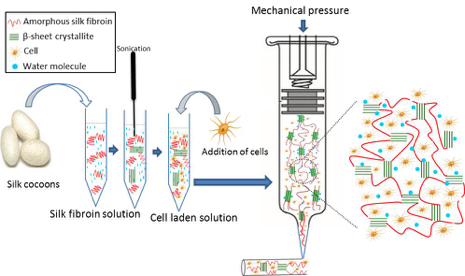
3D bio-printing of cell-laden scaffolds is a complex approach, which requires optimization of biomaterial fabrication, growth factor delivery and cell viability. Selection of the appropriate bioinks represents one of the main challenges in 3D-printing. Bioinks must not only be biocompatible and generate a mechanically stable 3D shape for the laden cells, but must also enable appropriate diffusion of nutrients, oxygen and metabolic waste products between the embedded cells and the culture medium. Silk fibroin (SF) has been increasingly recognized as a promising material for tissue engineering due to the ease of processing, its excellent biocompatibility and tuneable mechanical properties. Our group has strong expertise with the fabrication of SF scaffolds via the salt-leached method, which allows controllable pore size. These SF scaffolds were shown to support cell proliferation and formation of a mineralized extracellular matrix. 3D bio-printing in addition offers the advantage to directly include cells in the manufacturing process and to further control the architectural parameters such as pore structure and distribution, which are relevant for vascular ingrowth and osseo-integration of the scaffold in vivo.
This project focuses on the development of 3D cell-laden SF scaffolds, via the 3D bio-printing method, to promote bone regeneration. Our first aim is to develop a biocompatible 3D bioink, to investigate the cell-matrix interaction and the ability of cells to form a mineralized extracellular matrix within this 3D environment in vitro. Subsequently, the ability to promote bone regeneration in vivo will be evaluated.
Collaborators
Dr. Marina Rubert, ETH Zurich, Switzerland.
Acknowledgements
I gratefully acknowledge funding from the Overseas Doctoral Degree Program of the Chinese Scholarship Council.
Journal articles
Thimm, B. W., Wüst, S., Hofmann, S., Hagenmüller, H., & Müller, R. (2011). external pageInitial cell pre-cultivation can maximize ECM mineralization by human mesenchymal stem cells on silk fibroin scaffolds. Acta Biomaterialia, 7(5), 2218–2228. http://doi.org/10.1016/j.actbio.2011.02.004
Wüst, S., Godla, M. E., Müller, R., & Hofmann, S. (2014). external pageTunable hydrogel composite with two-step processing in combination with innovative hardware upgrade for cell-based three-dimensional bioprinting. Acta Biomaterialia, 10(2), 630–640. http://doi.org/10.1016/j.actbio.2013.10.016
Zhang, J., Zhao, S., Zhu, Y., Huang, Y., Zhu, M., Tao, C., & Zhang, C. (2014). external pageThree-dimensional printing of strontium-containing mesoporous bioactive glass scaffolds for bone regeneration. Acta Biomaterialia, 10(5), 2269–2281. http://doi.org/10.1016/j.actbio.2014.01.001