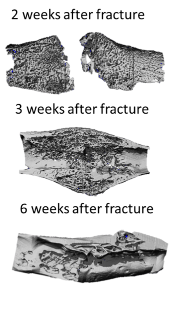
Quantitative Phenotyping of Bone Fracture Repair
Bone fracture healing is a highly complex process in which tissues are constantly replaced and different cell lineages are interacting. Such complexity and dynamism make the evaluation of resistance to refracture highly challenging in preclinical studies, since researchers are limited by the number of animals it is ethically, timely and financially possible to include in an investigation. This limitation led them to explore alternative ways to mechanical testing, which can require a large number of samples for providing an accurate assessment of the resistance to refracture. Some variables of the bone callus such as bone fraction and microarchitectural characteristics of the callus struts were already correlated to the resistance to refracture and are regularly reported in preclinical studies. However, other variant features which are likely to be predictor of callus strength are still not fully explored.
In recent years, researchers experimented emerging techniques for the assessment of fracture repair. Tools such as Raman spectroscopy, nanoindentation, scanning acoustic microscopy, in vivo micro-computed tomography (micro-CT), and high-resolution (HR) micro-CT have the potential to be adopted as new powerful instruments for the evaluation of the bone callus.
The aim of our research is to develop or adapt methods for a quantitative evaluation of the fracture callus. We also want to apply these techniques to preclinical study to better understand the effect of different treatments on fracture repair and demonstrate the relevance of a quantitative phenotyping of bone healing.
So far, we already developed or adapted a set of tools for estimating the resistance to refracture or to better understand the underlying processes. We implemented a new framework for quantifying the lacuno-canalicular network (LCN); we quantified bone material properties with Raman spectroscopy by using a novel method; we investigated and experimented indirectly the reliability of nanoindentation in a study on the anisotropy of the mouse femur; we inspected the effect of fixation on the bone tissue for future comprehensive studies on the bone callus; and we analyzed the microarchitecture of the bone callus with a new approach.

Collaborators:
Prof. Dr. David Little, The Children’s Hospital at Westmead, Australia.
Prof. Dr. Philipp Schneider and Prof. Dr. Aaron Schindeler, University of Southampton, UK.
Dr. Davide Carnelli, Dr. Diana Courty, and Tobias Keplinger, ETH Zurich, Switzerland.
Acknowledgments:
I would like to thanks the intern and students who worked under my supervision: Michael Pedroni (ETH), Bastien Chatton (ETH), Anna Balmelli (ETH), Janelle Herelle (MIT), Maria Skoura (University of Thessaloniki), Rowan Softley (University of Sheffield), Agnese Beretta-Piccoli (ETH), Marcel Thomas (ETH), Charlotte Bischoff (ETH), and Imke Fiedler (TU Hamburg).
Journal articels:
Casanova, M., Schindeler, A., Little, D., Müller, R., & Schneider, P. (2014). external page Quantitative phenotyping of bone fracture repair: a review. BoneKEy Reports, 3. http://doi.org/10.1038/bonekey.2014.45