Time-Lapsed Bone Response to Implantation in a Mouse Model of Disease and Treatment
Osteoporosis is prevalent among the elderly population and often causes devastating bone fractures, which requires long-term fixation with implants in order to restore the function of the fractured site. The long-term success of fracture fixation greatly depends on the early bone response following implantation, which regenerates the bone tissue at the implantation site to ultimately attain implant integration into the host bone. However, the poor bone quality and the compromised systemic conditions caused by osteoporosis may impair the early implant integration and jeopardize the long-term stability of the bone-implant system.
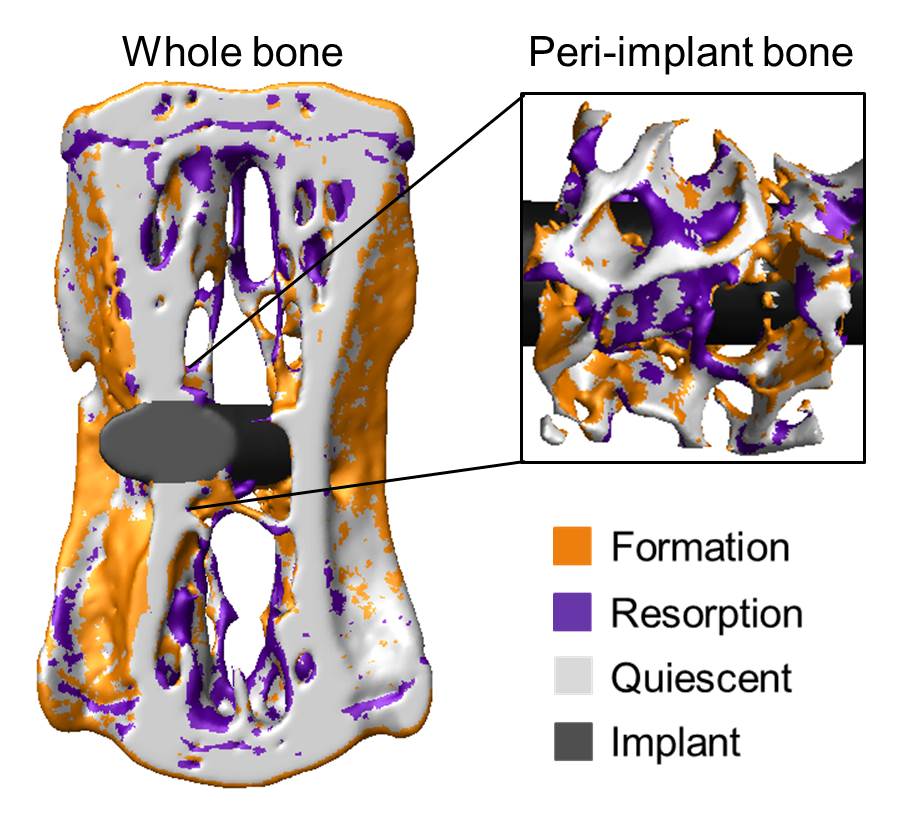
The bone remodeling process is a critical player in the complex biological response of bone following implant placement. During bone remodeling, in the peri-implant bone, damaged bone tissue is resorbed by osteoclasts and new bone is locally laid down by osteoblasts, leading to an adaptation in mass and structure of the peri-implant bone which is eventually responsible for the implant integration. Despite its importance, the process of bone remodeling following implantation and the consequential modifications in bone architecture remain poorly understood. Moreover, bone remodeling is mechanically controlled with the occurrence of bone formation being higher at sites with high strains and bone resorption being dominant at sites less strained. Therefore, controlled external mechanical loading has been proposed as a treatment to tune the process of bone remodeling in order to favor bone formation with the ultimate goal of enhancing implant integration. However, in order to develop effective loading protocols for osteoporotic fracture fixation, a more fundamental understanding of the interplay between mechanical loading and bone formation/resorption at the local tissue level in the peri-implant bone is mandatory.
The introduction of in vivo micro-computed tomography (micro-CT) and the latest development on image registration techniques provided access not only to the time evolution of changes in bone architecture, but also to the local identification and quantification of bone remodeling over time, including bone formation and bone resorption. However, the application of these techniques for the investigation of bone response around metallic implants used in clinics is limited by the marked image artifacts caused by the implant.
In this context, the main aim of this project was to develop a method allowing in vivo monitoring of the spatio-temporal changes in living bone following implantation, including bone remodeling and bone architecture. The method was then employed to characterize the response of osteoporotic bone and to answer the clinically relevant question whether mechanical loading could be used to improve peri-implant bone regeneration in osteoporotic bone.
Acknowledgments
We gratefully acknowledge funding from the Chinese Scholarship Council, the IOF-SERVIER Young Investigator Research Grant as well as the ECTS Postdoctoral Fellowship.
Journal articles
Li, Z., Kuhn, G., Salis-Soglio, von, M., Cooke, S. J., Schirmer, M., Müller, R., & Ruffoni, D. (2015). external pageIn vivo monitoring of bone architecture and remodeling after implant insertion: The different responses of cortical and trabecular bone.call_made Bone, 81, 468–477. http://doi.org/10.1016/j.bone.2015.08.017

