Micro-Computed Tomography: Applications and Services
Short Abstract: The technique of micro-computed tomography (micro-CT) provides three-dimensional images of internal structures of objects in the micrometre range non-destructively. The morphometry of these objects can be further evaluated by the application of image-processing algorithms to assess structural indices like object volume, surface, porosity, pore diameters and many more. Due to the non-destructive nature of micro-CT individual samples can be monitored longitudinally over time. Micro-CT is particularly used in the bone field to due to the excellent contrast of bone tissue to soft tissue, but it can be applied to many other structures as well.
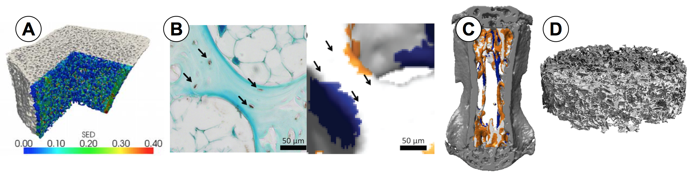
The on-going scientific projects conducted in our group are in some way all related with the application of micro-CT. Scans of bones from various species (human, murine, bovine etc.) provide the framework for most of the computational models developed in our group (figure A). In combination with histological images, these bone scans are used to gain spatial information of the gene expression of single bone cells (figure B). Scans of living mice (in vivo scans) allow us to study bone responses to mechanical loading in a longitudinal manner by repetitive scans. Thus, it is possible to see changes in bone microarchitecture and to define resorbed, formed or quiescent bone volumes (figure C). Micro-CT is also applied to investigate different aspects of isolated biological experiments (in vitro experiments) like the architecture of different substrate materials (scaffold) used for tissue engineering or the growth of mineralized tissue formed by cells over time (figure D).
With the application of micro-CT our lab aims to investigate the three-dimensional structural properties of various structures quantitatively and in a longitudinal manner where applicable. Due to the non-destructive nature of micro-CT we are able to use samples for further downstream applications like the implantation of a material into a bone defect or repeated scans of living organisms and cell cultures.
Contact
Institut für Biomechanik
Gloriastrasse 37/ 39
8092
Zürich
Switzerland