Bone Biomechanics: Understanding the Role of Bone Cells
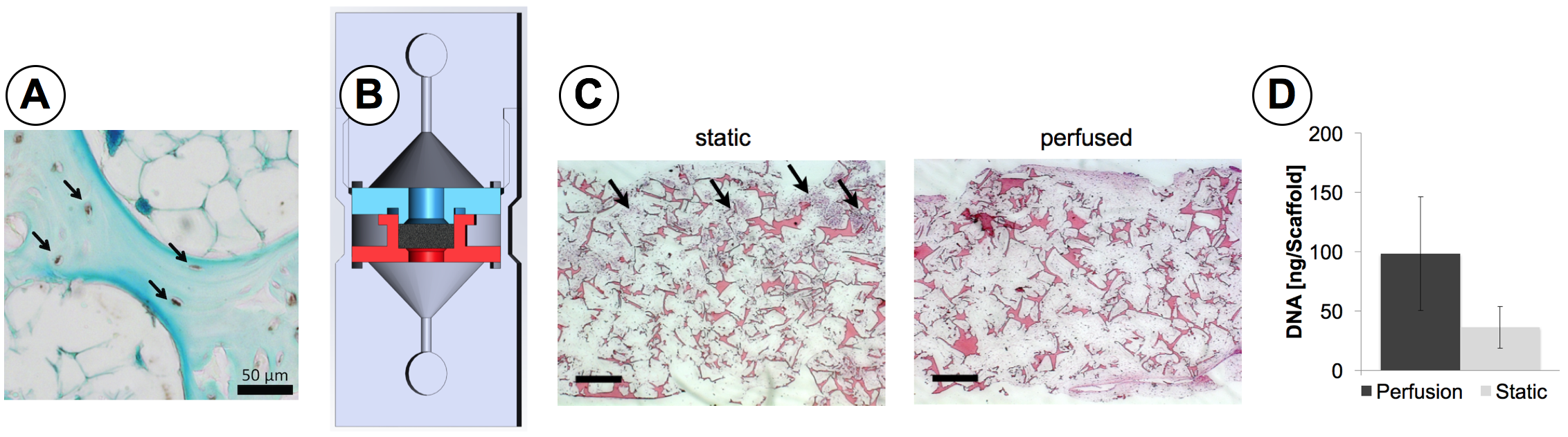
In the living organism (in vivo), bone cells are exposed to highly varying mechanical loads due to the irregular micro-architecture of bone. Our lab is therefore interested in quantifying gene expression locally. This is done by analysing the gene expression of individual cells or small cell populations from frozen histology sections (figure A). In isolated biological experiments (in vitro experiments), we aim to mimic the in vivo conditions of bone tissue to investigate the effect of the environment the cells are exposed to. Using different in-house designed bioreactor models we apply mechanical stimulation to cells (figure B). The effect of the stimulation on the cellular behaviour is then evaluated with histology (figure C), biomechanical assays (figure D), immunohistochemistry (figure A) or micro-computed tomography, just to name a few. Another interest of our in vitro studies is to evaluate the effect of the substrate material (scaffold) on which cells are cultured on. We are testing various scaffold designs, materials or production techniques, like 3D printing, using our bioreactor designs to assess the role of scaffolds in 3D cell cultures.
Taken all these experiments together, we aim to better understand the role of bone cells with respect to their biomechanical environment in vivo and in vitro. The evaluation of gene expression, enzymatic activity levels, cell numbers, microscopic anatomy of tissues or the amount and morphology of mineralized tissue helps us to interpret experimental outcomes by correlating results with the experimental design.